Topology-Preserving Coronary Artery Segmentation in CTA Volumes Using an nnFormer-CRF Model
DOI:
https://doi.org/10.64972/jiic.2026v4.471p15s:190-203Keywords:
Coronary CTA, Topology-Preserving Segmentation, nnFormer, Conditional Random Field, Vascular ConnectivityAbstract
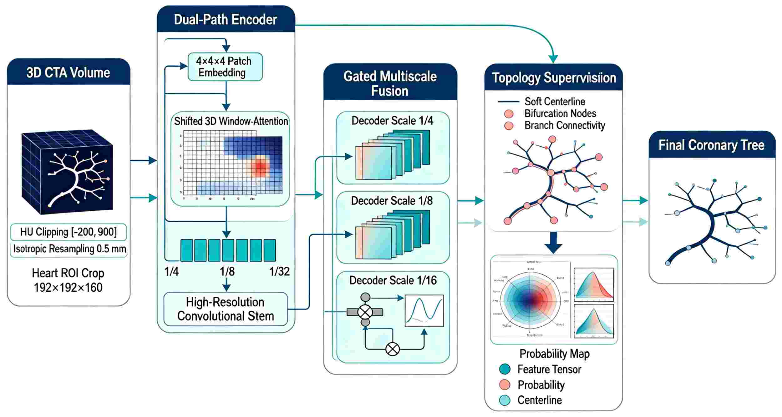
Coronary artery segmentation from CT angiography (CTA) is inherently difficult because the voxels of the distal vessels are small, there are changes in lumen contrast along the tree, and discontinuous or fused predictions occur due to calcified plaques. This paper develops a topology-preserving nnFormer-CRF model for full 3D coronary extraction. The encoder uses a moving volume window attention mechanism to capture long-range vascular context, and the topological branches adjust the soft centerlines and branch connections at three resolutions. Then, the graph conditional random field uses intensity, spatial, and tangential consistency potentials to fine-tune the logits, with these potentials constrained within an adaptive coronary region. To validate the model, a five-fold patient-level study was conducted, involving 420 CTA volumes from three centers. Its Dice coefficient is 0.912, the centerline Dice coefficient is 0.936, the mean symmetric surface distance is 0.31 mm, and the connection error per case is 0.71. Compared with the nnFormer backbone, the proposed method improved the recall of distal branches to 0.889 and reduced false vascular links by 34.8%. The best-case scenario of a large calcification had a Dice score of 0.79 after improvement. Inference for the resampled volume required 1.8s, and the refinement stage added an additional 0.23s. According to the results, Transformer context and structured anatomy refinement are complementary: the former can recover weak branches, while the latter avoids unreasonable shortcuts by reconstructing locally interrupted centerlines. The Framework offers good support for topology-sensitive coronary model building and quantitative CTA analysis.
References
Pan, L.-S., Li, C.-W., Su, S.-F., Tay, S.-Y., Tran, Q.-V., & Chan, W. P. (2021). Coronary artery segmentation under class imbalance using a U-Net based architecture on computed tomography angiography images. Scientific Reports, 11(1), 14493. https://doi.org/10.1038/s41598-021-93889-z
Baskaran, L., Al'Aref, S. J., Maliakal, G., Lee, B. C., Xu, Z., Choi, J. W., et al. (2020). Automatic segmentation of multiple cardiovascular structures from cardiac computed tomography angiography images using deep learning. PLOS ONE, 15(5), e0232573. https://doi.org/10.1371/journal.pone.0232573
Shin, C. I., Park, S. J., Kim, J. H., Yoon, Y. E., Park, E. A., Koo, B. K., & Lee, W. (2020). Coronary artery lumen segmentation using location–adaptive threshold in coronary computed tomographic angiography: A proof-of-concept. Korean journal of radiology, 22(5), 688. https://doi.org/10.3348/kjr.2020.0296
Li, Y., Wu, Y., He, J., Jiang, W., Wang, J., Peng, Y., et al. (2022). Automatic coronary artery segmentation and diagnosis of stenosis by deep learning based on computed tomographic coronary angiography. European Radiology, 32(9), 6037–6045. https://doi.org/10.1007/s00330-022-08761-z
Jávorszky, N., Homonnay, B., Gerstenblith, G., Bluemke, D., Kiss, P., Török, M., et al. (2022). Deep learning-based atherosclerotic coronary plaque segmentation on coronary CT angiography. European Radiology, 32(10), 7217–7226. https://doi.org/10.1007/s00330-022-08801-8
Pathak, A., Anwer, T., Verma, A., Alhujaily, M., Alahmari, M., Alshahrani, S., et al. (2026). Integrating precision medicine and artificial intelligence to prevent cardiotoxicity in cardiovascular drug therapy. Precision Medication, 3(1), Article 100078. https://doi.org/10.1016/j.prmedi.2026.100078
Lin, A., Kolossváry, M., Motwani, M., Išgum, I., Maurovich-Horvat, P., Slomka, P. J., & Dey, D. (2022). Artificial intelligence in cardiovascular imaging for risk stratification in coronary artery disease. Radiology: Cardiothoracic Imaging, 4(1), e210108. https://doi.org/10.1148/ryct.210108
Karimi, D., Dou, H., & Gholipour, A. (2022). Medical image segmentation using transformer networks. IEEE Access, 10, 29322–29332. https://doi.org/10.1109/ACCESS.2022.3156894
Shit, S., Paetzold, J. C., Sekuboyina, A., Ezhov, I., Unger, A., Zhylka, A., Pluim, J. P. W., Bauer, U., & Menze, B. H. (2021). clDice—A novel topology-preserving loss function for tubular structure segmentation. 2021 IEEE/CVF Conference on Computer Vision and Pattern Recognition, 16555–16564. https://doi.org/10.1109/CVPR46437.2021.01629
Hatamizadeh, A., Tang, Y., Nath, V., Yang, D., Myronenko, A., Landman, B., Roth, H. R., & Xu, D. (2022). UNETR: Transformers for 3D medical image segmentation. 2022 IEEE/CVF Winter Conference on Applications of Computer Vision, 574–584. https://doi.org/10.1109/WACV51458.2022.00069
Gao, Y., Zhou, M., & Metaxas, D. N. (2021). UTNet: A hybrid transformer architecture for medical image segmentation. Medical Image Computing and Computer Assisted Intervention—MICCAI 2021, 12903, 61–71. https://doi.org/10.1007/978-3-030-87199-4_6
Shi, Y., Zhang, J., Ling, T., Lu, J., Zheng, Y., Yu, Q., Qi, L., & Gao, Y. (2021). Inconsistency-aware uncertainty estimation for semi-supervised medical image segmentation. IEEE Transactions on Medical Imaging, 41(3), 608–620. https://doi.org/10.1109/TMI.2021.3117888
Luo, X., Wang, G., Liao, W., Chen, J., Chen, Y., Song, T., et al. (2022). Semi-supervised medical image segmentation via uncertainty rectified pyramid consistency. Medical Image Analysis, 80, 102517. https://doi.org/10.1016/j.media.2022.102517
Xiao, Z., Su, Y., Deng, Z., & Zhang, W. (2022). Efficient combination of CNN and transformer for dual-teacher uncertainty-guided semi-supervised medical image segmentation. Computer Methods and Programs in Biomedicine, 226, 107099. https://doi.org/10.1016/j.cmpb.2022.107099
Malhotra, P., Gupta, S., Koundal, D., Zaguia, A., & Enbeyle, W. (2022). Deep neural networks for medical image segmentation. Journal of Healthcare Engineering, 2022, 9580991. https://doi.org/10.1155/2022/9580991
Isensee, F., Jaeger, P. F., Kohl, S. A. A., Petersen, J., & Maier-Hein, K. H. (2021). nnU-Net: A self-configuring method for deep learning-based biomedical image segmentation. Nature Methods, 18(2), 203–211. https://doi.org/10.1038/s41592-020-01008-z
Bertels, J., Eelbode, T., Berman, M., Vandermeulen, D., Maes, F., Bisschops, R., & Blaschko, M. B. (2021). Theoretical analysis and experimental validation of volume bias of soft Dice optimized segmentation maps in the context of inherent uncertainty. Medical Image Analysis, 67, 101833. https://doi.org/10.1016/j.media.2020.101833
Mehrtash, A., Wells, W. M., Tempany, C. M., Abolmaesumi, P., & Kapur, T. (2020). Confidence calibration and predictive uncertainty estimation for deep medical image segmentation. IEEE Transactions on Medical Imaging, 39(12), 3868–3878. https://doi.org/10.1109/TMI.2020.3006437
Jungo, A., Meier, R., Ermis, E., Blatti-Moreno, M., Herrmann, E., Wiest, R., & Reyes, M. (2020). On the effect of inter-observer variability for a reliable estimation of uncertainty of medical image segmentation. Medical Image Analysis, 64, 101718. https://doi.org/10.1016/j.media.2020.101718
Zhang, D., Tai, X.-C., & Lui, L. M. (2021). Topology- and convexity-preserving image segmentation based on image registration. Applied Mathematical Modelling, 100, 218–239. https://doi.org/10.1016/j.apm.2021.08.017
Zeleznik, R., Foldyna, B., Eslami, P., Weiss, J., Alexander, I., Taron, J., et al. (2021). Deep convolutional neural networks to predict cardiovascular risk from computed tomography. Nature Communications, 12(1), 715. https://doi.org/10.1038/s41467-021-20966-2
Lin, A., Manral, N., McElhinney, P., Killekar, A., Matsumoto, H., Kwiecinski, J., et al. (2022). Deep learning-enabled coronary CT angiography for plaque and stenosis quantification and cardiac risk prediction: An international multicentre study. The Lancet Digital Health, 4(4), e256–e265. https://doi.org/10.1016/S2589-7500(22)00022-X
Karimi, D., & Salcudean, S. E. (2020). Reducing the Hausdorff distance in medical image segmentation with convolutional neural networks. IEEE Transactions on Medical Imaging, 39(2), 499–513. https://doi.org/10.1109/TMI.2019.2930068
Ma, J., Chen, J., Ng, M., Huang, R., Li, Y., Li, C., Yang, X., & Martel, A. L. (2021). Loss odyssey in medical image segmentation. Medical Image Analysis, 71, 102035. https://doi.org/10.1016/j.media.2021.102035
Huang, H., Lin, L., Tong, R., Hu, H., Zhang, Q., Iwamoto, Y., ... & Wu, J. (2020, May). Unet 3+: A full-scale connected unet for medical image segmentation. In ICASSP 2020-2020 IEEE international conference on acoustics, speech and signal processing (ICASSP) (pp. 1055-1059). Ieee. https://doi.org/10.1109/ICASSP40776.2020.9053405
Wang, W., Chen, C., Ding, M., Yu, H., Zha, S., & Li, J. (2021). TransBTS: Multimodal brain tumor segmentation using transformer. Medical Image Computing and Computer Assisted Intervention—MICCAI 2021, 12901, 109–119. https://doi.org/10.1007/978-3-030-87193-2_11
Liu, Z., Lin, Y., Cao, Y., Hu, H., Wei, Y., Zhang, Z., Lin, S., & Guo, B. (2021). Swin transformer: Hierarchical vision transformer using shifted windows. 2021 IEEE/CVF International Conference on Computer Vision, 9992–10002. https://doi.org/10.1109/ICCV48922.2021.00986
Karimi, D., Dou, H., Warfield, S. K., & Gholipour, A. (2020). Deep learning with noisy labels: Exploring techniques and remedies in medical image analysis. Medical Image Analysis, 65, 101759. https://doi.org/10.1016/j.media.2020.101759
Cai, L., Gao, J., & Zhao, D. (2020). A review of the application of deep learning in medical image classification and segmentation. Annals of Translational Medicine, 8(11), 713. https://doi.org/10.21037/atm.2020.02.44
Ahmad, H. M., Khan, M. J., Yousaf, A., Ghuffar, S., & Khurshid, K. (2020). Deep learning: A breakthrough in medical imaging. Current Medical Imaging, 16(8), 946–956. https://doi.org/10.2174/1573405615666191219100824
Downloads
Published
How to Cite
Issue
Section
License
Copyright (c) 2026 Ivan Jelen, Dalibor Sedláček

This work is licensed under a Creative Commons Attribution-NonCommercial-ShareAlike 4.0 International License.